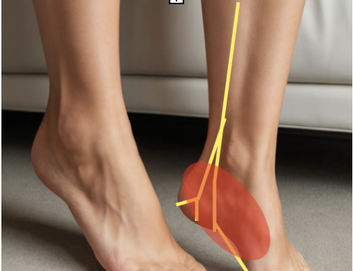
Calcaneofibular Ligament: Lateral Ankle Ligament Injury (CFL and ATFL)
The calcaneofibular Ligament band connects the calcaneus (calcaneofibular) to the lateral aspect of the fibula. The peroneus longus and brevis tendon cover the ligament. The calcaneofibular ligament and the posterior and anterior talofibular ligaments resist inversion ankle sprains. Special tests that can detect injuries include MSKUS (ultrasound), palpation, and orthopedic ankle tests. A severe calcaneofibular ligament injury can lead to ankle instability.
CFL and AFTL
The calcaneofibular ligament injury rarely occurs without also injuring that anterior talofibular ligament. During an ankle sprain, the anterior talofibular ligament comes to tension first due to its anterior location, and then once the AFTL fails, the calcaneofibular ligament becomes injured. An ankle sprain that causes damage to both the calcaneofibular ligament and the anterior talofibular ligament is considered a severe lateral ankle sprain and can cause ankle instability.
Instability
This form of ankle instability involves talocural joint and the inability to control talar tilt. This form of ankle instability can cause the ankle to become stiff, swollen, painful, and lead to a sensation of the ankle giving out. Chronic ankle instability causes repeated ankle sprains and ankle tendonitis, most commonly posterior tibial tendonitis.
Diagnosis
Calcaneofibular ligament orthopedic tests include the talar tilt test, anterior drawer test, and tenderness with palpation of the ankle ligament.
Talar Tilt Test– Foot in neutral, and doctor create a tilting force to invert the ankle to test the integrity of the calcaneofibular ligament. Laxity and increased movement with pain compared to the uninjured ankle are positive and CFL injuries.
Anterior Drawer Test– Foot in neutral, and doctor create an anterior shear force on the ankle ligaments. Increased movement with pain compared in the uninjured ankle is considered a positive and a CFL injury.
Palpation– Place finger just below the tip of the fibula on the inferior posterior aspect and work down to the calcaneus bone. Tenderness to palpation 4-5 days after injury with positive talar tilt and anterior drawer tests is predictive of a lateral ligament rupture.
Special Tests include MRI, X-Ray, and MSK ultrasound used to confirm a diagnosis of calcaneofibular ligament injury or ankle instability.
Our editorial practices include evidence-based practices, interventions, and recommendations.

GET IN TOUCH WITH DR. DEAN
YOU should be able to move the way you’d like to move without experiencing pain. YOU should be able to experience freedom and energy knowing there’s nothing holding back from giving your life 110%. Dr. Dean would like to learn more about your challenges with a quick phone or email before beginning treatment. Contact him today.